This month, fMRI brain imaging celebrates its 20th anniversary. And so it should. It has come to dominate cognitive neuroscience.
Massive amounts of precious funding are poured into it and thousands of studies are published every year.
Critics say it’s not worth it – brain imaging will not explain the mind. But this criticism is premature: brain imaging is maturing and holds explanatory promise.
Fun and games
Lying in the MR scanner, you are playing rock, paper, scissors on a computer screen. Half the time, you’re told your opponent is a computer program, the rest of the time that it’s another person.
Though the sequence of play is, in fact, exactly the same, the game feels different when you believe you are playing against a person.
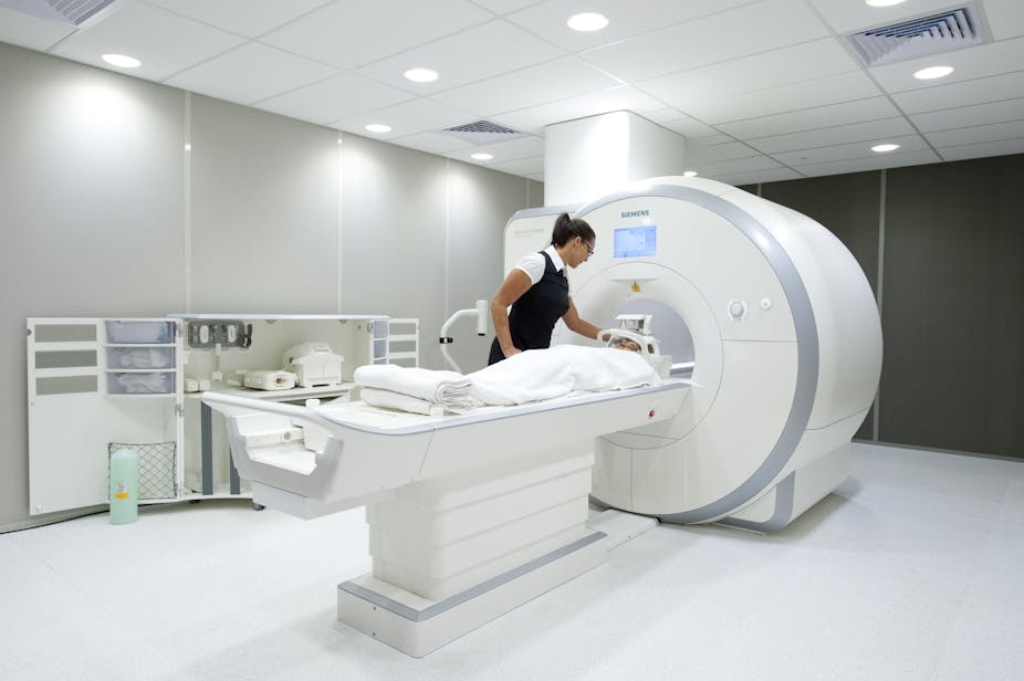
This a functional magnetic resonance imaging (fMRI) experiment. As you play, a massive magnetic field pulsates through your body and picks up minute changes in the blood flow of your brain.
The pattern of changes differs when you believe you play a person as opposed to when you believe your opponent is a computer.
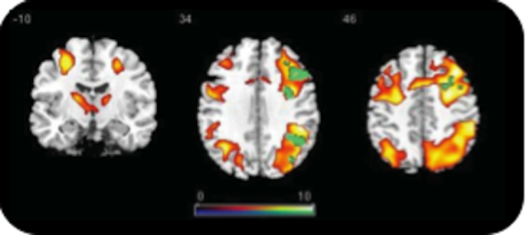
Subtract the patterns from each other, do some more statistics, and the result is a neat coloured blob on a picture of the brain.
MRI brain scan from JonO on Vimeo.
Since the only real difference between the playing conditions is your mental state, the difference in brain activity must be specific to that type of mental state.
So the blob signifies the part of the brain that explains why we interact differently with people and computers.
Meaningful blobs
The technique for this research – functional magnetic resonance imaging – was published 20 years ago this month in Science. It caused a dramatic shift in neuroscience.
Thousands of papers have subsequently been published, all with images of blobs on brains.
All sorts of mental phenomena are now routinely scanned, from viewing simple shapes to emotional states, such as disgust, religious belief, love and beyond, to psychosis, depression and psychopathy.
We love the blobs and have become accustomed to them. No matter what the quality of the message, if it’s accompanied by images of brain blobs and explained with some neurobabble, we are ready to believe it.
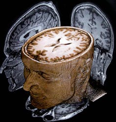
The blobs, we are told, and believe, signify activity that explains the mind. But they actually do no such thing.
Blobs on brains are generally boring, misused and misunderstood. But understood in the right light, they are also exciting and will help us understand the mind.
It is boring, for instance, to confirm that when the mind changes, the brain changes. After all, the two are one. What would truly have been amazing is if fMRI had revealed changes in the mind are not accompanied by brain changes.
The exciting thing, in fact, is how brain activity changes.
Early promise vs reality
Before neuroimaging, 100 years of neurological research had used stroke patients and soldiers wounded by shrapnel to indirectly infer which part of the brain underpins which mental function.
Then it suddenly seemed everything we had learned could be evaluated directly with fMRI. We thought we would explain the mind in no time.
But reality quickly curbed this enthusiasm: fMRI doesn’t directly measure brain activity at all.
The MR scanner is a big pulsating magnet that can track minute changes in blood flow and blood flow is not thought, belief or emotion. It’s not even brain activity as such.
So what is fMRI data evidence for? Research in the last ten years has begun to answer this question.
Neuroscientists are now generally satisfied that fMRI reliably tracks important activity in the neurons of the brain’s grey matter. This activity is some kind of combination of neurons being modulated by other neurons and neurons that fire.
Statistical contortions
Findings from fMRI are indirect in another way.
Going from the magnetic pulses to the coloured blobs is fraught with methodological and statistical choices that can completely change what the blobs will look like.
That’s because mental states and brain areas do not have a one-to-one relationship. And all the variability this causes generates scepticism.
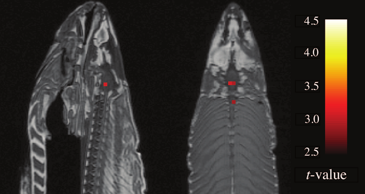
How could it not when a dead salmon put in a scanner can, via judicious statistical choices, also generate neat coloured blobs?
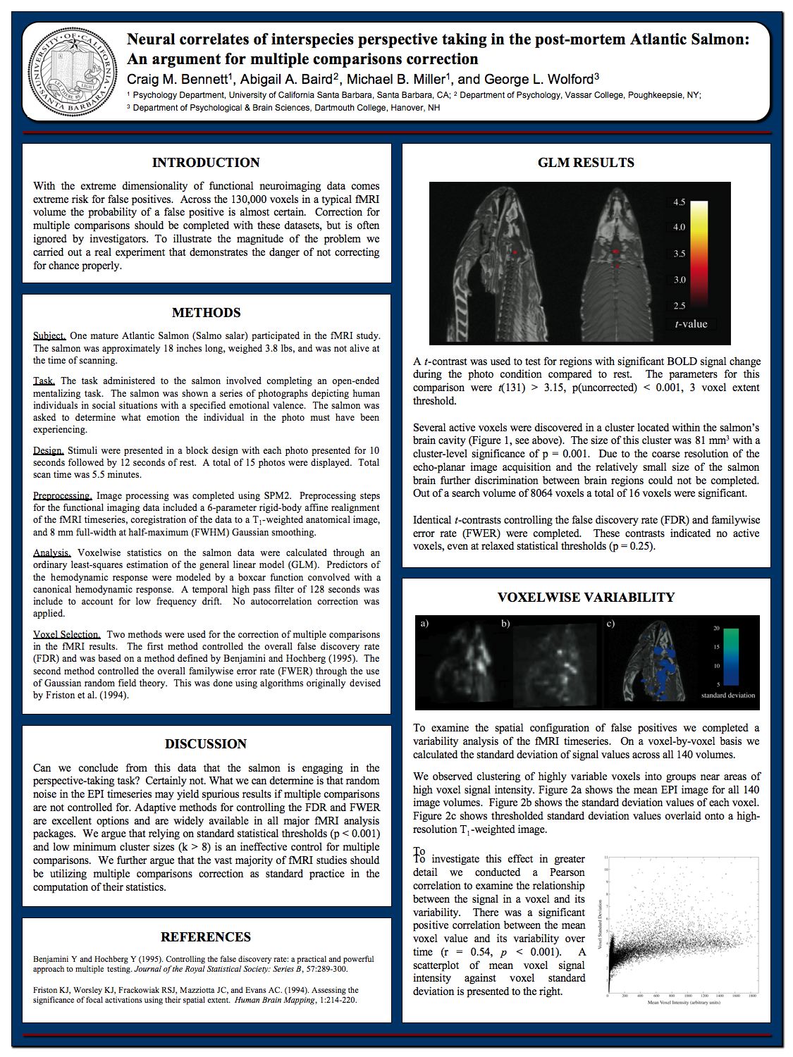
But stable patterns of blobs are evident and a lot of the variability seems due to subtle differences in tasks and experimental designs. Nevertheless, there’s still serious discussion about the best method of brain scanning and no doubt many findings from the first 20 years of fMRI will not survive the next 20.
Even so, people are still asking what the point of getting an fMRI map of brain activity is. After all, a map in itself doesn’t explain anything.
And is all the funding put into brain imaging worth it, if blobs are all we get?
This scepticism is premature – it’s normal for an emerging science to begin by mapping out the landscape.
Slowly, hypotheses about the connecting mechanisms will emerge. These hypotheses, in turn, will help build better maps that generate even better hypotheses about the underlying mechanisms.
Beyond blobology
The first 20 years of fMRI has helped build a pretty detailed map of the brain. The next phase is finding the causal mechanisms and the principles behind them.
The aim is to go beyond mere blobology.
On the basis of the brain map, researchers have begun to formulate possible models of brain mechanisms – which blobs cause which other blobs.
The time-course of brain activity allows us to rule out some mechanisms and rule in others. This way, we learn how mental states are built up, emerge from sensory input, and how they lead to behaviour.
In other words, the blobs don’t explain the mind, but their causal interconnections and the underlying statistical analysis may.
The scepticism about neuroimaging is also based on limited assumptions. Understanding the brain just by doing fMRI scanning is too hard, just as it’s too hard to get a full interactive understanding of city life merely by mapping it from a plane flying above.
Many different techniques are required to complement each other.
The electrical signals of the brain, for instance, are measured with EEG, which gives great temporal resolution that combines well with the spatial resolution of fMRI. And there are numerous other exciting ways of looking at the brain.
A further, essential method for discovering causal mechanisms is trying to break them and see what happens, just as a theory of traffic flow can be tested by interrupting the flow.
The fMRI map can guide interventions where transcranial magnetic stimulation (temporarily) breaks brain mechanisms.
The co-discoverer of DNA, Francis Crick, famously said, “you’re nothing but a pack of neurons”.
In the next 20 years fMRI will, in spite of its shortcomings, help us illustrate this in great detail.